OTHER METHODS OF VISION CORRECTION

JASNIJE SUTRA POČINJE DANAS.

Content of the article

Text author: Dr sc. Bojan Kozomara, dr med.

ABOUT PHAKIC INTRAOCULAR LENSES

If the diopter is too high or the corneal tissue is too thin, it does not mean that the diopter cannot be corrected.

Indeed, the implantation of phakic intraocular lenses represents the best and most effective alternative to laser vision correction.

fakicna intraokularna sociva en

WHAT ARE PHAKIC INTRAOCULAR LENSES?

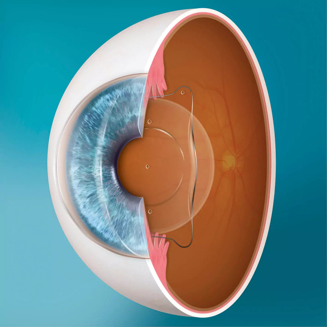
Phakic intraocular lenses are intraocular implants made of a special type of collamer, a biocompatible material that is fully adaptable to the human eye. Currently, the most popular implant of this type is the Visian ICL (Implantable Collamer Lens) phakic intraocular lens.

intraocular lenses, ophthalmologist

WHAT ARE THE BASIC ADVANTAGES OF THIS LENS?

– It can correct a wide range of diopters from -3.00 D to -23.00 D, and from +0.50 D to +10.00 D.

– It effectively corrects astigmatism up to +6.00 D. It is easily implanted behind the iris and in front of the natural crystalline lens without the need for fixation to surrounding eye tissues.

– Its use reduces trauma to surrounding tissues, shortens the procedure, and accelerates postoperative recovery.

– The diameter of the optical part of the lens is significantly larger, making it an ideal implant for patients with naturally wide pupils.

– With the implantation of this lens, the eye’s ability to accommodate, providing clear vision at both near and far distances, is not lost. Therefore, it is often recommended for young individuals who do not need reading glasses.

– Lens implantation is reversible; the lenses can be explanted, and the condition can be restored to its original state if the patient desires for any reason.

HOW DOES THE SURGICAL PROCEDURE LOOK LIKE?

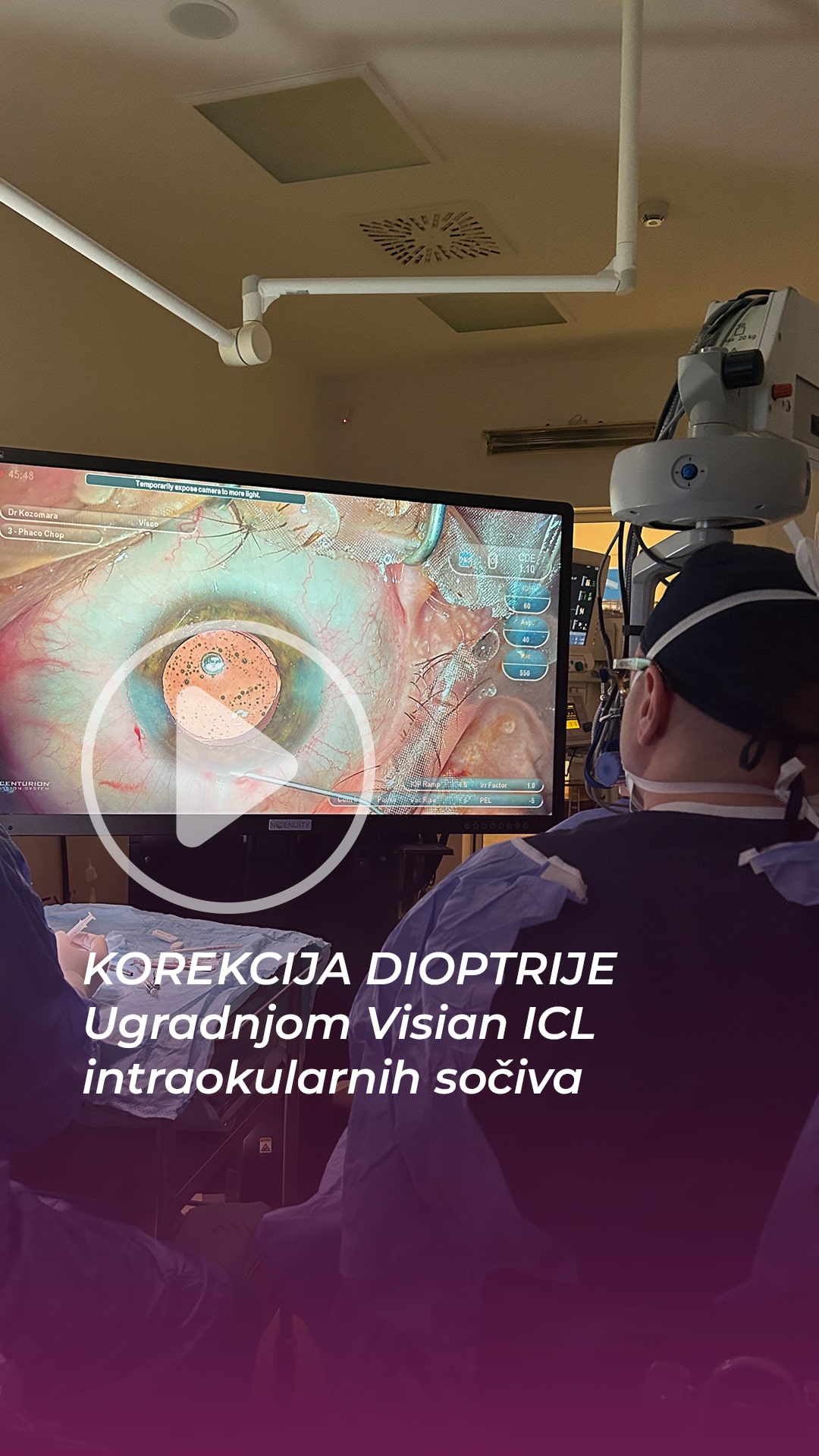
The implantation is simple and painless, involving the insertion of the lens through a corneal incision of just a few millimeters. The lens is first implanted into the anterior chamber of the eye and then carefully positioned behind the iris and in front of the natural crystalline lens. The corneal incision heals on its own, eliminating the need for sutures. The surgical procedure is performed under topical anesthesia and lasts only 10 minutes per eye.

WHAT DOES THE POSTOPERATIVE PERIOD LOOK LIKE?

The patient leaves the hospital immediately after the procedure. Vision may be blurred for several days, during which antibiotic drops are instilled in both eyes. The patient can return to their daily activities after just 5 days following the surgery.

VIDEO

laserska korekcija dioptrije, dr kozomara
Copyright ©2024 all rights reserved
Skip to content