Personalized treatment of the retina
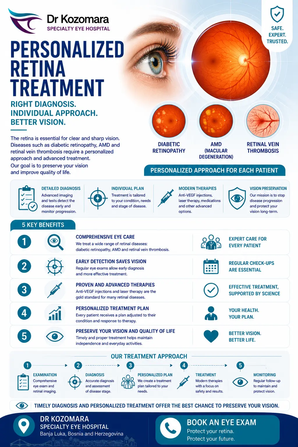
The retina is one of the eye's most important structures, and its diseases are among the most complex in ophthalmology. That's why we take a personalized approach — from diabetic retinal disease to AMD to vein thrombosis, each condition gets a treatment tailored to the individual patient.
The retina is one of the most important structures of the eye, and its diseases are among the most complex in ophthalmology. That is why we take a personalized approach — for each condition we select a treatment tailored to the individual patient, with the aim of preserving and restoring vision.
Conditions we treat
Diabetic retinal disease
In people with diabetes, a circulatory disturbance in the retinal capillary network leads to blood-vessel damage and haemorrhage. Patients report weaker, poorer vision and dark spots or "floaters" in their field of vision. Read more on our diabetic retinopathy page. We advise all people with diabetes to have at least one ophthalmic examination per year, regardless of symptoms.
Age-related macular degeneration (AMD)
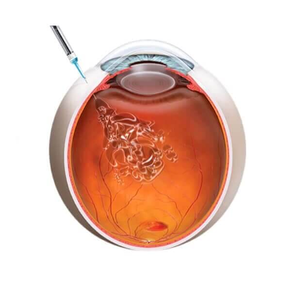
In this disease central vision weakens while peripheral vision remains preserved. There are two forms: the dry form (gradual development, accumulation of drusen; peripheral vision remains preserved) and the wet form (a sudden, aggressive form with bleeding in the macula). The wet form, if detected in time, is successfully treated with anti-VEGF therapy (Avastin, Lucentis, Eylea), while late detection and scarring reduce the effect of treatment.
Retinal vein thrombosis and arterial embolism
Also known as an "eye infarction", it occurs when a retinal blood vessel becomes blocked — most often due to poorly controlled blood pressure or elevated blood lipids — and can cause partial or complete loss of part of the visual field. It is treated with anti-VEGF therapy and laser photocoagulation, along with a mandatory internal-medicine and neurology evaluation to prevent cardiac and cerebral complications.
Diagnosis of retinal diseases
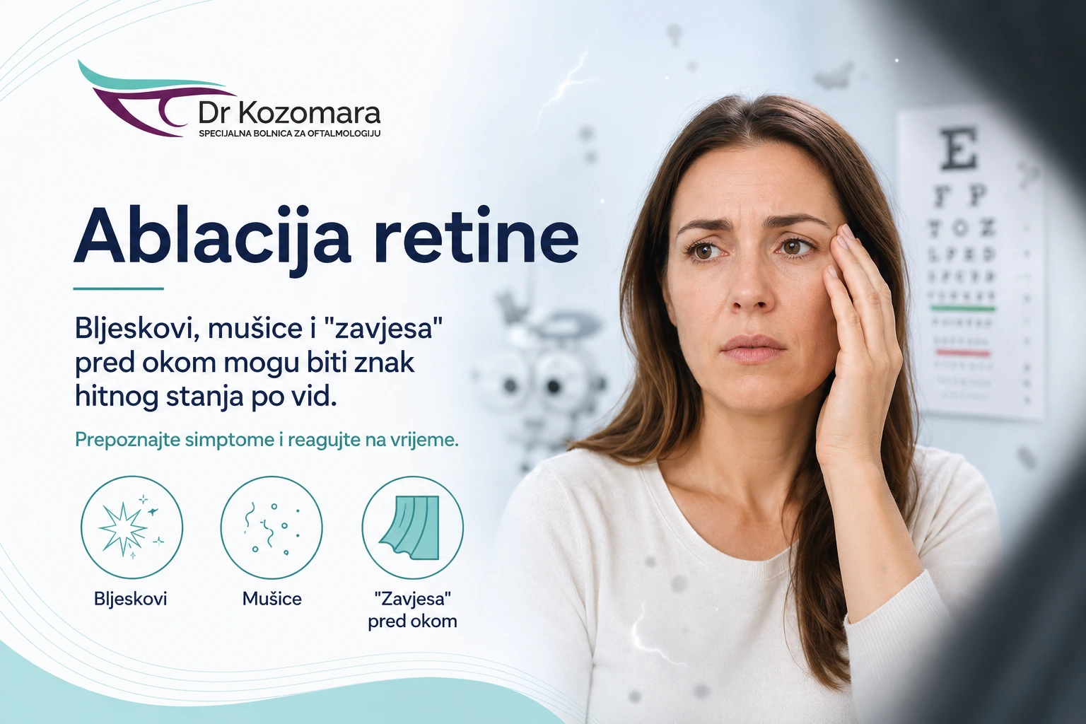
To make a precise diagnosis we use ultrasound diagnostics, OCT (optical coherence tomography), fundus photography and electroretinography. Sudden changes — flashes, a "curtain" or a shower of new floaters — may indicate retinal detachment and require an urgent examination.
Noticing a shadow, floaters or weakening central vision? Book a fundus examination at Dr Kozomara Hospital.
Everything you need to know about personalized treatment of the retina
Retinal treatment diagnosis
Diseases of the posterior segment of the eye, the retina, are often the most difficult to treat in modern ophthalmology. To determine the state of the eye’s retina, we use several methods:
- Ultrasound diagnostics
- OCT – optical coherence tomography
- Fundus photography
- Electroretinographic analysis
Diseases of the posterior eye segment
This is also the most common and complex eye damage in diabetic patients. Due to changes in metabolism, there is a disturbance in circulation in the capillary network of the retina, leading to damage to arteries and veins and leakage of blood onto the surface of the retina as well as into the vitreous body. Patients describe these damages as a decrease in visual acuity and poorer image quality, as well as the appearance of “dark spots or floaters” in the visual field. If you are diabetic, we recommend that you undergo an ophthalmological examination at least once a year, regardless of whether you recognize any of the symptoms mentioned above.
Age-related macular degeneration
Age brings many new, beautiful experiences, but also takes something from the overall health picture of a person. We know that one of the organs that can feel the effects of aging is precisely the eye, more specifically the macula, which is one of the most important anatomical parts of each eye.
How does this disease manifest?
Visual acuity decreases, and the damage is particularly pronounced in the “central” vision. The patient feels a large “spot” in front of one or both eyes, causing objects to appear blurry, but still maintains preserved peripheral vision.
Types of age-related macular degeneration
In practice, two forms of this condition are present: dry and wet. In the dry form of age-related macular degeneration, damage to central vision occurs gradually, over several years, even decades. Due to pronounced atherosclerosis of the retinal blood vessels, there is accumulation of cellular metabolic waste, hindering the normal functioning of remaining living cells. The accumulated material is called drusen.
The wet form is much more aggressive, occurring suddenly, marked by the appearance of a distinctly dark “spot” in front of the eye. Sudden vision loss is caused by the rupture of atherosclerotic retinal blood vessels and bleeding into the area of the macula. Even with this form of macular degeneration, peripheral vision may remain preserved.
How is it treated?
The dry form of age-related macular degeneration still cannot be treated, but the patient will never completely lose vision. Specifically, they will always retain what is known as peripheral vision, or vision outside the macula.
The wet form of senile macular degeneration is successfully treated if detected early with the application of anti-VEGF therapy (Avastin, Lucentis, Eylea). However, if detected late or inadequately treated, therapy may not be very effective due to the rapid formation of scarring in the macula, which remains untreatable.
Thrombosis of the vein and arterial embolism
This condition is also known as an eye infarction and often occurs as a result of uncontrolled or poorly controlled blood pressure, as well as high levels of fat in the blood.
Just as in a heart or brain infarction, a blockage of an eye artery or vein by a clot can occur, consequently leading to damage to a part of the visual field or complete visual acuity.
How is it treated?
Vein thrombosis and arterial embolism can be treated with the application of anti-VEGF therapy (Avastin, Lucentis, Eylea) or laser photocoagulation of the damaged areas on the retina.
In addition to ophthalmological treatment, it is necessary to perform detailed internal medicine and neurological examinations to prevent more severe damage to the heart or brain.
Gallery
Learn how other people overcame their dependence, and find out how you can, too
Join thousands who have achieved freedom from glasses and contacts
FAQs and information about vision correction
Everything you need to know about our practice and procedures